
Thoracic arachnoid web myelopathy
I met a patient recently who had an interesting condition I’d not come across before. First, a bit about his history:
 He was a gentleman around 60 years old who had deteriorating lower limb symptoms for the last 10 years. These included weakness, sensory changes and spasticity. He’d been to several doctors and claimed he usually felt “fobbed off”. They did scan his lumbar spine which didn’t show any abnormalities. He was fit and healthy prior to his symptoms and regularly played tennis. As it progressed he got to the point he was struggling to walk. As his symptoms continued to worsen he met a different doctor who really listened to him. He ordered a full spine MRI which showed he had a thoracic web myelopathy.
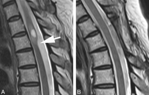
What is a thoracic arachnoid web myelopathy?
We all have a layer of translucent tissue around our spinal cord called the arachnoid membrane. It’s fibrous appearance is thought to look like a spiders web, hence the name arachnoid web. In very rare cases you can get a thickening of the arachnoid layer which can compress the spinal cord. This can affect how electrical signals are transmitted down to your lower limbs. This can cause symptoms such as altered reflexes, weakness, sensory changes, spasticity, altered gait and neuropathic pain. The symptoms you get depend on the level and severity of the spinal cord compression. All functions of the spinal cord above the level of compression remain unimpaired.